
Profile
Team
Equipment
Publications
Terms of use
Contact |
Profile
Cryogenic electron microscopy (cryo-EM) has revolutionised structural studies in the biological and chemical sciences in recent years. This method uses samples prepared by vitrification, i.e. rapid immersion in a solution of liquefied cryogen (ethane or propane) at a temperature below -140°C. Such a process prevents the formation of crystalline ice and is currently the best possible way to preserve the native structure of the sample under study, which is compatible with high-resolution electron-beam imaging, unlike the heavy metal staining and resin embedding methods used in the past.
The creators of the cryo-EM technique were honored with the 2017 Nobel Prize in Chemistry, and since then, the popularity of the method has grown exponentially and many research centers around the world are investing in cryo-EM-related specialist laboratories (core facilities).
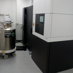
The 200kV Glacios microscope operates at CeNT as part of the Cryomicroscopy and Electron Diffraction Core Facility, which, in addition to the microscope itself, is fully equipped for sample preparation and vitrification and with a comprehensive IT infrastructure dedicated to the storage and processing of large amounts of data. There are currently 2 centers in Poland carrying out cryo-EM research (CeNT UW and Solaris UJ in Krakow), but only on the microscope at CeNT is research being carried out covering all three application streams of the technology:
- single Particle Analysis – for structural studies of purified macromolecules e.g. proteins, nucleic acids, viruses with a resolution of less than 3Å
- cryo-tomography (cryo-ET) – for 3D imaging of unique structures on a macromolecular scale, e.g. liposomes, secretory vesicles (EVs), cubosomes, etc.
- electron microdiffraction (micro-ED) – for determining the spatial structure of nanocrystals of small organic molecules, minerals, MOFs, proteins, etc. At CeNT, we have implemented this technique as the first center in Poland and one of several in Europe.
|
|
Equipment
| Glacios 200kV cryo-EM microscope
Equipped with:
- Schottky field emission source
- Ceta D 200kV CCD camera
- Falcon 3EC 200kV direct electron detection camera
- Phase plate solution for enhanced contrast
- Autoloader holding up to 12 grids in a cassette
- Micro-ED package
- TEM tomography data acquisition software
- EPU software for single particle analysis experiments
- EPU-D for micro-ED data collection
|
|
| Vitrobot for automatic vitrification of biological samples
Glow discharge unit (PELCO easiGlow, Ted Pella) for glow discharging TEM grids
Auxillary equipment for micro-ED sample preparation (sonicator, vacuum chamber)
IT infrastructure for data storage & analysis including storage infrastructure, servers, computational clusters, software |
|
|
|
Selected publications
- Tian Y, Liang R, Kumar A, Szwedziak P, Viles JH. 3D-visualization of amyloid-β oligomer interactions with lipid membranes by cryo-electron tomography. Chem Sci. 2021 Mar 31;12(20):6896-6907. doi: 10.1039/d0sc06426b. PMID: 34123318; PMCID: PMC8153238.
- Alvarez-Malmagro J, Jablonowska E, Nazaruk E, Szwedziak P, Bilewicz R. How do lipid nanocarriers – Cubosomes affect electrochemical properties of DMPC bilayers deposited on gold (111) electrodes? Bioelectrochemistry. 2020 Aug;134:107516. doi: 10.1016/j.bioelechem.2020.107516. Epub 2020 Mar 20. PMID: 32222670.
- Alvarez-Malmagro J, Matyszewska D, Nazaruk E, Szwedziak P, Bilewicz R. PM-IRRAS Study on the Effect of Phytantriol-Based Cubosomes on DMPC Bilayers as Model Lipid Membranes. Langmuir. 2019 Dec 17;35(50):16650-16660. doi: 10.1021/acs.langmuir.9b02974. Epub 2019 Dec 5. PMID: 31746606.
- Cytryniak A, Nazaruk E, Bilewicz R, Górzyńska E, Żelechowska-Matysiak K, Walczak R, Mames A, Bilewicz A, Majkowska-Pilip A. Lipidic Cubic-Phase Nanoparticles (Cubosomes) Loaded with Doxorubicin and Labeled with 177Lu as a Potential Tool for Combined Chemo and Internal Radiotherapy for Cancers. Nanomaterials (Basel). 2020 „Nov 16;10(11):2272. doi: 10.3390/nano10112272. PMID: 33207760; PMCID: PMC7696353.”
- Piszczatowska K, Czerwaty K, Cyran AM, Fiedler M, Ludwig N, Brzost J, Szczepański MJ. The Emerging Role of Small Extracellular Vesicles in Inflammatory Airway Diseases. Diagnostics (Basel). 2021 Feb 2;11(2):222. doi: 10.3390/diagnostics11020222. PMID: 33540806; PMCID: PMC7913078.
|